Dental disease
 Dental disease is the most common disease of small animals. The most common types of dental disease among small animals include periodontal (gum) disease, endodontic (nerve) disease, and tooth resorption, where part or all of a tooth is lost because body cells attack the tooth.
Cavities or decay are not as frequent in dogs as they are in humans, and apparently do not affect cats.
Even though dental disease can be severe and painful, and seriously affect the general health status and quality of life of an individual, animals usually conceal their discomfort, so clinical signs and symptoms can be very subtle. Possible signs of dental disease include bad breath, excessive drooling, pawing at the face, difficulty eating, bleeding or recessed gums, and tooth loss.
Dental disease can be suspected based on reported symptoms and a conscious oral and physical examination. However, a definitive diagnosis and treatment plan can only be established with the animal under general anesthesia; the standard of care for the diagnosis of dental disease includes dental probing and charting, and full-mouth x-rays. Once a diagnosis is established, treatment is usually performed while the animal remains under anesthesia.
Common treatment for canine and feline dental disease includes ultrasonic scaling and polishing of teeth above and below the gumline, periodontal surgery, root canal therapy, extractions and fillings.
When properly diagnosed, most dental conditions are treatable and the prognosis is excellent. Adequately treated patients usually experience relief and owners normally notice a dramatic improvement in attitude, appetite and overall health.
Oral and Maxillofacial Surgery
Cats and dogs also present with diseases and conditions that require oral or maxillofacial surgery. The most common conditions include traumatic injuries, such as fractures or lacerations; palatal defects, such as oronasal fistula and cleft palate; and oral tumors.
Many patients with traumatic injuries are initially treated by the Emergency and Critical Care Service for initial stabilization, and are then transferred to the Dentistry and Oral Surgery Service for specific maxillofacial procedures. To diagnose their injuries, typically x-rays and/or CT scanning are performed.
The treatment approach and techniques differ with each case and depend on a variety of factors. The outcome and prognosis of most maxillofacial traumatic injuries is good.
Patients with oral tumors typically require an initial appointment where we conduct an examination and imaging scans to determine the exact nature of the tumor and any associated disease. After a precise diagnosis is made, many oral tumors can be removed surgically. Often times, the Oncology Service will recommend additional treatments such as chemotherapy or radiation. The outcome and prognosis of patients with oral tumors varies, and depends on the exact nature of the tumor and stage of the disease.
Patients with palatal defects typically undergo definitive surgical repair after a planning session that may include CT imaging. Depending on the size and location of the defect, final correction may require a multi-stage approach. The outcome of palatal defect repair is usually good.
Maxillofacial Reconstruction for "Bella" Dasson
 Bella is a dog that was involved in a serious car accident when she was 11 months old. Even though her owners did not witness the accident and were not there to help her immediately, Bella had the strength and courage to make it back home on her own. As soon as her owners realized what had happened, they took Bella to a local emergency clinic, where she was stabilized and referred to Cornell’s Emergency and Critical Care Service. When she arrived at our hospital, she had multiple maxillofacial injuries, as well as a fractured right front limb.
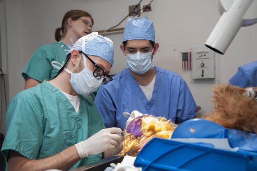
The next day, we performed a CT scan of Bella's head to better document the extent and severity of the injuries sustained to her face. Bella’s upper and lower jaws had multiple, severe fractures. Without surgery, she would have been permanently disfigured and disabled. Bella was transferred to the Dentistry and Oral Surgery Service for repair of her maxillofacial injuries and facial reconstruction. Dr. Santiago Peralta performed the surgery assisted by Shantell Hassey, a fourth-year veterinary student.
The procedure took a total of 7 hours to complete; the results could not have been better, however. We were able to stabilize her fractures and properly align her bones during the surgery, our orthopedic service also casted Bella’s fractured right front limb. After surgery we monitored Bella in our Intensive Care Unit overnight.
Bella recovered incredibly well and was discharged to her family the day after surgery. Although we placed a feeding tube during surgery in case she was not able to eat on her own while her injuries healed, Bella started eating without assistance within the first 24 hours after surgery. Bella required one more surgical procedure a few weeks after her maxillofacial injuries healed to address some dental injuries. Today, Bella is completely healthy, both physically and emotionally, and is enjoying her life to the fullest.
The American Veterinary Dental College
The American Veterinary Dental College is recognized as the specialist certification organization in veterinary dentistry in North America by the American Board of Veterinary Specialties. AVDC diplomats are veterinary dental specialists.
American Veterinary Dental Society
The American Veterinary Dental Society is a non-profit organizations created to advance the knowledge, education, and awareness of veterinary dentistry and increase awareness of the importance of this facet of animal medicine.
Veterinary Oral Health Council
The Veterinary Oral Health Council is an independent organization that awards a registered seal to products intended to help retard plaque and tartar on the teeth of animals.


 Dental disease is the most common disease of small animals. The most common types of dental disease among small animals include periodontal (gum) disease, endodontic (nerve) disease, and tooth resorption, where part or all of a tooth is lost because body cells attack the tooth.
  Bella is a dog that was involved in a serious car accident when she was 11 months old. Even though her owners did not witness the accident and were not there to help her immediately, Bella had the strength and courage to make it back home on her own. As soon as her owners realized what had happened, they took Bella to a local emergency clinic, where she was stabilized and referred to Cornell’s Emergency and Critical Care Service. When she arrived at our hospital, she had multiple maxillofacial injuries, as well as a fractured right front limb.