Study unlocks potential for early-intervention arthritis therapies
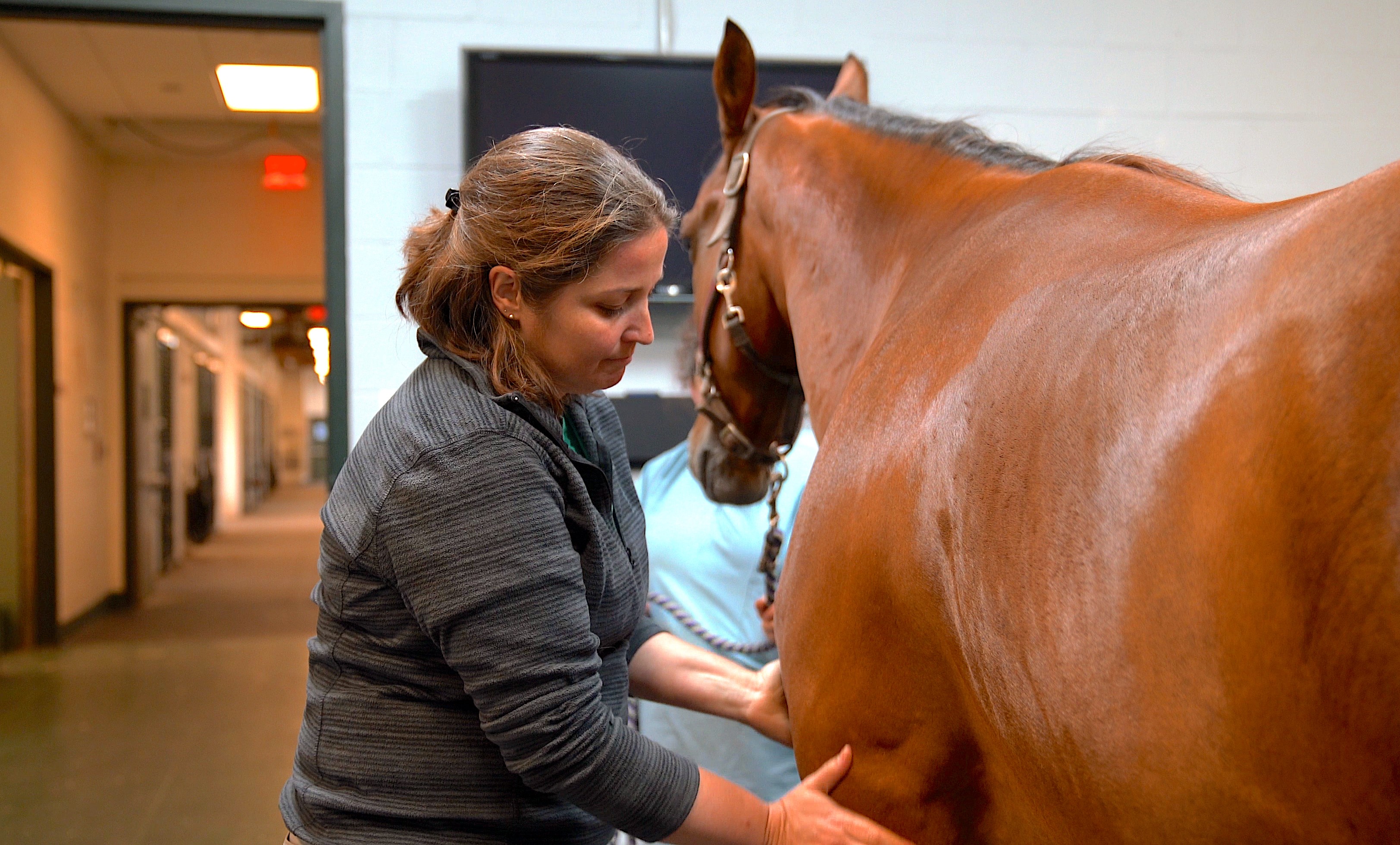
A new way to diagnose and treat arthritis at an early stage could be on the horizon, according to a study by a Cornell team led by Michelle Delco, D.V.M. ’02, Ph.D. ’16, the Harry M. Zweig Assistant Research Professor in Equine Health, and an equine surgeon at the Cornell University Hospital for Animals and Cornell Ruffian Equine Specialists in the Department of Clinical Sciences.
Arthritis is caused by degradation of cartilage in joints. When this damage occurs following an injury, such as a fracture or severe ankle sprain, it is known as post-traumatic osteoarthritis, or PTOA.
At least five-and-a-half million Americans suffer from PTOA, and it is common in both human and equine athletes.
“Arthritis is one of the top causes of chronic pain and disability in people and in horses,” says Delco. “And PTOA is probably a lot more common than is reported or recognized.”
Evidence suggests that early intervention could be the key to preventing PTOA. But detecting the disease at the initial stages has long been a struggle. And once it has taken root, the disease cannot be reversed.
“If you and I both sprain our ankle really badly,” says Delco, “there is no way to predict which one of us will end up with arthritis. We can't see or test for the subtle beginnings of cartilage damage, so there's no evidence that we need to treat anything.”
Symptoms of PTOA, such as joint pain or loss of mobility, tend only to appear years or decades after injury. MRIs can pick up the disease, but only after structural problems in the cartilage have already developed.

This means that, once PTOA is diagnosed, it is often too late to turn back the clock. The critical question is how to diagnose the disease before it has become irreversible.
Delco and her team began looking for clues that would allow the disease to be picked up before serious cartilage damage has set in. “We were looking for a marker that we can measure, near the time of injury, that can predict whether a patient is likely to develop arthritis down the road, and therefore, who needs treatment.”
Their results, in a recent paper published in Osteoarthritis and Cartilage, reveal the existence of a marker that is highly associated with early joint injury. This indicator can be measured at the nanoscale, at the level of DNA.
The key lies in analyzing the subcellular mechanisms that cause arthritis.
Following a joint injury, the mitochondria, the energy factories of the cell, stop working as they should. This mitochondrial dysfunction triggers many downstream events that lead, ultimately, to irreversible joint disease.
If only you could tell whether the mitochondria were dysfunctional, Delco thought, you might detect the onset of arthritis before it was irreversible. You could also stop the disease in its tracks with early interventions.
Delco’s lab cultivated cartilage cells to model the onset of arthritis. They noticed that the cells were releasing mitochondrial DNA into the surrounding fluid in response to stress.
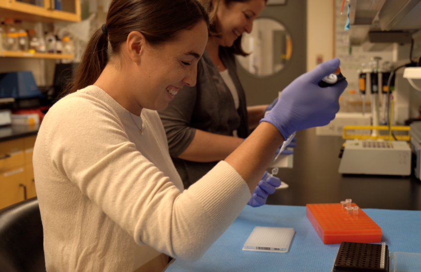
The presence of mitochondrial DNA in the fluid, detected using a PCR test, was a good indicator that the mitochondria had stopped working properly.
Further experiments revealed that after injury to a joint surface, mitochondrial DNA leaks into the synovial fluid, the thick liquid that lubricates the joints. It was a warning signal of the very beginnings of arthritis.
“Our study shows that when mitochondrial dysfunction happens in cells within the cartilage, mitochondrial DNA is released. If we sample the joint fluid, we can detect that mitochondrial DNA. And the more mitochondrial DNA in the fluid, the more likely the patient is to have cartilage damage and early arthritis.”
The discovery unlocks the possibility of new, early-intervention therapies.
Delco’s lab had previously shown that mitochondria in cartilage cells could be protected by treating with a peptide drug called SS-31, after cartilage injury.
Without an understanding of whether mitochondrial disfunction is occurring, though, it is impossible to know when that treatment should be applied. And without an indicator of faulty mitochondria, there is no way to tell if the treatment is working.
Delco’s discovery helps to resolve this problem. In the study, some horses with impact injuries were treated with SS-31, while others were not. The horses who had been treated had less mitochondrial DNA in their synovial fluid compared with the untreated group. This indicated that the treatment was effective.
More recently, Delco and her team have started analyzing synovial fluid from humans with knee injuries. They have found high levels of mitochondrial DNA in the joint fluid. “We have much more work to do,” says Delco, “but early data suggest this is likely true across species.”
Written by Tomas Weber


