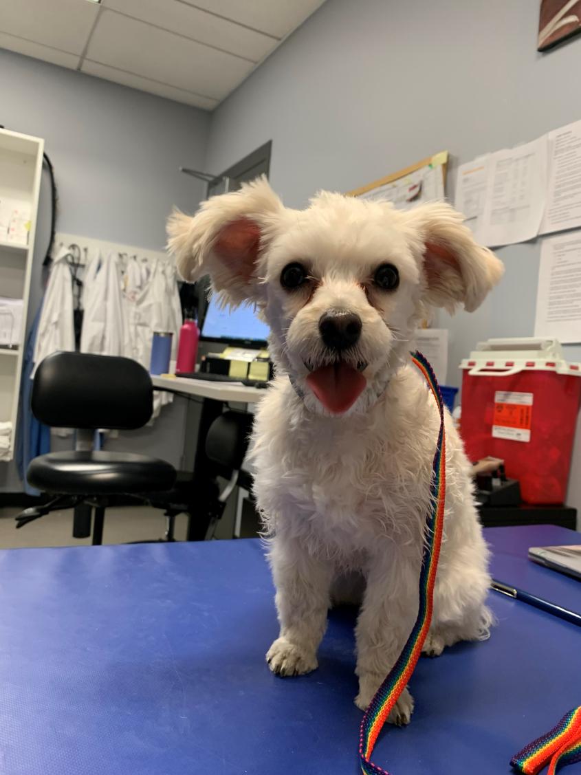
Tiny dog’s big heart procedure is first of its kind at Cornell
When a patient is only 6.4 pounds, certain procedures can get tricky. That was the case for Buttercup, an 11-month-old Morkie (Maltese-yorkie mix), who suffered from a deadly congenital cardiac defect. At most hospitals, Buttercup would have had to go through a more invasive and painful surgery to fix her condition, but the cardiology team at the Cornell University Hospital for Animals performed a highly unusual procedure to keep her at her healthiest.
According to Buttercup’s owners, the tiny pup had a heart murmur since she was born. While she had good energy at home, she would begin coughing if she got excited or exercised. They brought Buttercup to Cornell, where the cardiology team diagnosed her with patent ductus arteriosus (PDA), the most common congenital cardiac defect in dogs.
“All dogs have a ‘ductus arteriosus’ (DA) in utero,” says cardiology resident Dr. Christophe Bourguignon. “It is a vessel allowing normal blood oxygenation inside their mom as they do not have functional lungs to breathe and use the placenta to do so. A few minutes/hours after birth, this vessel closes, allowing the lungs to take over completely. Occasionally, some dogs’ DA does not close and stay ‘patent,’ hence the term PDA.” While the DA is open, this vessel allows a massive amount of blood to overflow the lungs and the heart. “Most dogs will eventually go into congestive heart failure and die,” Bourguignon adds. “Drugs do not work well, if at all.”
Race against time
With Buttercup’s diagnosis confirmed, the cardiology team knew they had to act fast. “The vessel needs to be closed as soon as possible to prevent the complication of heart failure,” says Dr. Romain Pariaut, associate professor and section chief of cardiology. “This condition causes a very characteristic heart murmur that any veterinarian should be able to recognize with a stethoscope at the time of a puppy’s visit.”
To fix the PDA, clinicians typically do a catheter-based occlusion of the PDA, which uses long catheters inserted into a small artery in the patient’s leg and guiding the catheter to the DA using via fluoroscopy and echocardiography to view the process. Then, they place a plug inside the vessel to close the hole and stop the blood flow. However, Buttercup was so small that this traditional method wasn’t feasible — her femoral artery was too small even for the smallest catheter available.
A pup in luck
The traditional alternative for Buttercup would have been a more invasive procedure in which the chest is open and a surgeon manually closes the PDA. But Buttercup was in luck: “The nice and unusual thing with Buttercup is that we offered to modify the catheter-based occlusion technique in order to access the PDA without having to open her chest,” says Bourguignon.
That modified approach involves threading the catheter through Buttercup’s femoral vein instead — which is typically a little bigger and more elastic than the artery and has a lower blood pressure and a lower risk of bleeding. “This different approach had not yet been done at Cornell,” says Pariaut, who had performed it one time previously at a different institution.
This technique is much trickier — the catheter must be guided inside and through the heart’s chambers first before it can reach the pulmonary artery, and cardiologists must use a small flexible wire to probe the wall of the artery until they find the opening of the PDA. An additional difficulty is that the hole through which the clinicians must drop the coil is much smaller and harder to target.
The Cornell cardiology team embarked on this unusual procedure, partnering with the anesthesia and pain management service. “No cardiac procedure would be possible at Cornell without the anesthesia team’s expertise,” says Bourguignon. “For me, the anesthesiologist is as important as the cardiologist.”
Assisted by Bourguignon and with the support of the cardiology technicians, Pariaut took on the main task of guiding the catheter through the femoral vein and closing the DA with the coil. “He is a world-renowned expert on minimally-invasive cardiac procedures,” notes cardiology colleague Bruce Kornreich, D.V.M. ’92, Ph.D. ’05, senior extension associate and director of the Cornell Feline Health Center. In just under two hours, Pariaut and Bourguignon successfully closed the PDA in Buttercup. “We were able to show that we can do this other, more complicated technique to fix PDAs in smaller patients,” says Pariaut.
A condition cured
After her surgery, Buttercup recovered well. At the time of her scheduled recheck three months after the procedure, cardiology resident Dr. Lawrence Santistevan confirmed by echocardiography that the procedure had been successful and the PDA was completely closed.
PDA is one of the few cardiac conditions that can be cured if diagnosed and treated early, says Pariaut. “After treatment, the animals have a completely normal life. The procedure is very safe, and we can offer it to a wide range of dog sizes. PDAs are also easy to diagnose — it is very important that they make sure that a veterinarian listens carefully to their pet’s heart whenever they get a new puppy.”
Written by Lauren Cahoon Roberts


