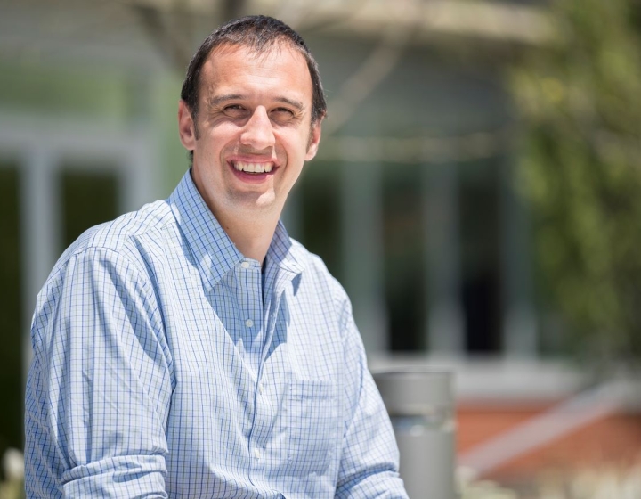
Embryologist wins scientific achievement award for research on how heads form
 Pugs and collies; horses and people— their faces are amazingly different, yet during early embryonic stages all look very similar. Studying how young freewheeling cells in embryos move around then eventually settle down to establish this fundamental pattern has helped Dr. Drew Noden, professor of embryology and animal development at the College of Veterinary Medicine, unlock several secrets about how faces acquire their unique yet highly recognizable forms.
In recognition of his contributions, Noden was named the recipient of the 2014 Henry Gray/Lippincott Williams Wilkins Scientific Achievement Award by the American Association of Anatomists (AAA). Named after Henry Gray, the author of the well-known human anatomy book Gray’s Anatomy, first published in 1858 and now in its 40th edition, the award is the AAA's highest scientific honor. It recognizes unique and meritorious contributions to anatomical sciences and achievements in this field by its most distinguished members. As the recipient, Noden will present a lecture at the AAA Annual Meeting in April 2014. The award includes a $1,500 honorarium and travel reimbursement to receive the medal at the AAA Banquet.
Noden has focused his career studying how the many disparate building blocks of the head are directed to form highly conserved musculoskeletal systems such as jaws and faces. While many in the field of development specialize in specific tissue types, Noden took a more comprehensive approach, studying several different tissue types, including the peripheral nerves, skeletal structures, blood vessels, and muscles, and creating intricate maps of how their progenitors moved about inside the head at different developmental stages.
 “Most people tend to focus on one tissue type—the tooth, the jaw, or a specific type of muscle,” said Noden. “My lab’s approach was different because we studied many types at once, thereby creating a more global picture of how the head forms. First, we produced maps of where all these tissues come from, carefully mapping the movements of the cells that build them. Then we explored the underlying mechanisms by surgically changing the relationships between different cell populations to better understand how they communicate with each other prior to forming specialized assemblies of tissues.”
Before cells in the developing embryo settle on their final role, they move around as groups or individual progenitor cells. Some move in ordered patterns, others in apparent chaos. Noden mapped their movements by labeling newly-formed progenitor cells, then watching where they would go. By then examining how cells moved and differentiated when challenged by different neighboring cells, the nature of communications among them was revealed.
“If you’re a cell wandering around chaotically, at some point you need to settle down and get a life,” said Noden. “Part of what determines the choices each cell makes has to do with the influence of other cells they meet at different times along the way. It is much like life."
“Working in the Veterinary College where our patients present with an enormous variety of facial anatomies, and trying to figure out how embryos generate all these variations on a common plan have been very rewarding. As I’ve always told my students-- if you spend enough time with living embryos, eventually one will whisper a secret to you. That secret could be one that no one else knows, but that embryos have held for hundreds of millions of years. I couldn’t imagine anything more exciting and also humbling.”


